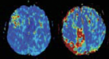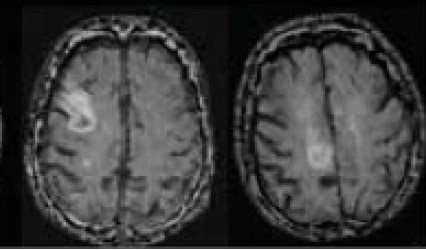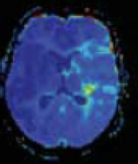


Explanation of Perfusion Imaging and Its Importance
Perfusion imaging is a crucial technique in medical imaging that measures the flow of blood through the brain’s vascular network. It provides vital information about the delivery of oxygen and nutrients to brain tissue, which is essential for maintaining healthy brain function. By visualizing and quantifying blood flow, perfusion imaging helps in diagnosing and treating various neurological conditions, including stroke, brain tumors, and traumatic brain injuries.
Perfusion imaging is particularly important in acute settings, such as during a stroke, where timely and accurate information about blood flow can significantly impact treatment decisions and outcomes. It helps clinicians identify areas of the brain that are at risk due to reduced blood flow (ischemia) and differentiate between regions that are permanently damaged and those that can potentially be saved with appropriate intervention.
Relative Cerebral Blood Flow (rCBF)
Relative Cerebral Blood Volume (rCBV)
Mean Transit Time (MTT)
Time to Maximum (Tmax)
Capillary Transit-Time Heterogeneity (CTH)
Oxygen Extraction Fraction (OEF)
Cerebral Metabolic Rate of Oxygen (CMRO2)
Benefits of Microvascular Modeling in Diagnosis and Treatment Planning
Microvascular modeling offers significant advantages in the diagnosis and treatment planning of various neurological conditions. Here are some key benefits:
Enhanced Diagnostic Accuracy
Early Detection of Disease
Personalized Treatment Planning
Monitoring Treatment Efficacy
Improved Prognostication
Cercare Medical’s advanced microvascular modeling technology leverages these unique metrics to provide a comprehensive and detailed assessment of brain health. By going beyond traditional perfusion imaging, we empower clinicians with the tools needed to make more informed decisions, improve patient care, and ultimately enhance clinical outcomes

Dynamic Contrast Enhanced (DCE) MRI provides detailed information about tissue vascularity by tracking the distribution of a contrast agent over time. It is particularly useful in oncology for assessing tumor vascularity and permeability.
Efficiency and Precision: The software automatically processes DCE MRI data, providing quick and accurate results, thus enhancing clinical workflow efficiency.
Model-Free Perfusion Markers
Comprehensive Analysis: Includes model-free perfusion markers such as relative Cerebral Blood Flow (rCBF), essential for evaluating tissue perfusion without the need for complex models.
Extended Tofts Model
Advanced Metrics: Utilizes the widespread Extended Tofts model to calculate important parameters like Ktrans (volume transfer constant), Ve (extravascular extracellular space), and Vp (plasma volume), offering detailed insights into capillary permeability and tissue vascularity.
Cercare Medical’s DSC Perfusion (MRI) module is designed to deliver detailed cerebral perfusion analysis, aiding in the diagnosis and treatment planning for various neurological conditions.
Fully Automated Processing
Efficiency and Reliability: Automatically detects and processes DSC MRI data, ensuring rapid and consistent results. This automation minimizes manual intervention to the point where no manual interaction is necessary, reducing the risk of human error and streamlining clinical workflows.
Model-Free Perfusion Markers
Essential Data: Provides crucial model-free perfusion markers such as relative Cerebral Blood Flow (rCBF) and relative Cerebral Blood Volume (rCBV), which are vital for assessing brain perfusion without the need for complex models.
Microvascular Modeling
Advanced Insights: Incorporates microvascular modeling to deliver detailed metrics like Capillary Transit-Time Heterogeneity (CTH) and model-based Oxygen Extraction Fraction (OEF). These advanced markers provide deeper insights into the cerebral microvasculature, enhancing diagnostic precision.
Contrast Agent Extravasation Correction
Accurate Measurements: Corrects for the extravasation of contrast agents, ensuring the accuracy of perfusion measurements. This feature is crucial for obtaining accurate cerebral perfusion biomarkers such as blood flow or blood volume, free from artifacts caused by contrast leakage.
By integrating these cutting-edge features, Cercare Medical’s DSC Perfusion (MRI) module empowers clinicians with the tools needed for precise and comprehensive cerebral perfusion analysis, ultimately improving patient care and outcomes.
Cercare Medical’s Diffusion Weighted Imaging (DWI) module is designed to enhance the detection and characterization of acute ischemic stroke and other conditions affecting water diffusion in the brain.
Fully Automated Processing
Streamlined Workflow: Automatically detects and processes DWI data, ensuring accurate and swift results. This automation reduces the need for manual intervention, increasing efficiency and consistency in clinical practice.
Mean Diffusion Weighted Image (DWI) and Apparent Diffusion Coefficient (ADC)
Critical Metrics: Calculates mean DWI and ADC maps, which are essential for identifying areas of restricted diffusion often indicative of acute stroke or tumors. These maps provide crucial information for diagnosing and planning treatment for various brain pathologies.
Optional Correction for Magnetic Field Inhomogeneities
Enhanced Accuracy: Includes an option for magnetic field inhomogeneity correction to improve the accuracy of diffusion measurements, ensuring reliable data for clinical decision-making.
By integrating these advanced features, Cercare Medical’s Diffusion Weighted Imaging (DWI) module empowers clinicians with the tools needed for precise diffusion analysis and ensures alignment between diffusion images and other images processed through Cercare Medical Neurosuite through native image alignment (co-registration), ultimately assisting in improvement of patient care and outcomes.
CT Perfusion (CTP) is a technique used to evaluate cerebral blood flow, blood volume, and other perfusion metrics by monitoring the passage of an iodine-containing contrast agent through the brain using CT imaging.
• Fully Automated Processing: Cercare Medical’s software automates the detection and processing of CTP data, ensuring consistent and rapid delivery of perfusion images in the PACS. The images are fully compliant with the DICOM standard and can be viewed alongside primary imaging sequences. Generally, it can be thought of as perfusion without the hazzle.
• Perfusion Markers: The software provides standard perfusion markers such as relative cerebral blood flow (rCBF), relative cerebral blood volume (rCBV), and mean transit time (MTT). These markers help in assessing the extent of perfusion abnormalities.
• Advanced Metrics: In addition to standard markers, the software offers advanced metrics like CTH and model-based OEF, providing a comprehensive view of cerebral hemodynamics .
• Integration with Clinical Workflow: The vendor neutral and automated processes allow seamless integration with clinical workflows, facilitating quick and efficient diagnosis and treatment planning.
The relative cerebral blood flow. SVD* & PARAMETRIC
MR DSC, MR
DCE*, CTP
The relative cerebral blood volume. SVD & PARAMETRIC
MR DSC, CTP
Mean transit time of the passage
of blood through a voxel. SVD &
Parametric.
MR DSC, CTP
Capillary Transit-time heterogeneity. A measure of the dispersion in intra-voxel capillary transit times.
MR DSC, CTP
Model-based oxygen extraction fraction
MR DSC, CTP
The relative model-based
cerebral metabolic rate of oxygen
MR DSC, CTP
Delay from site of measurement
of the arterial input function
concentration time-curve and site of measurement of the tissue
concentration-time curve.
MR DSC, MR DCE, CTP
The timepoint at which the
residue function attains its
maximum value.
MR DSC, CTP
Measure of the lack of information. I.e. a value larger than 0.05 means that there is very little information in the particular voxel.
MR DSC, CTP
Coefficient of Variance also referred to as relative transit time heterogeneity ‘RTH’ in scientific and clinical literature.
MR DSC, CTP
Extravasation of contrast agent in a particular voxel, i.e. leakage from the vascular compartment to the extravascular compartment.
Time to peak of the concentration-time curve.
Note that the TTP map is not related to the SVD or parametric deconvolution methods.
MR DSC, MR DCE, CTP
Time from peak until wash out of contrast agent - MR DSC
.
MR DSC
Cli
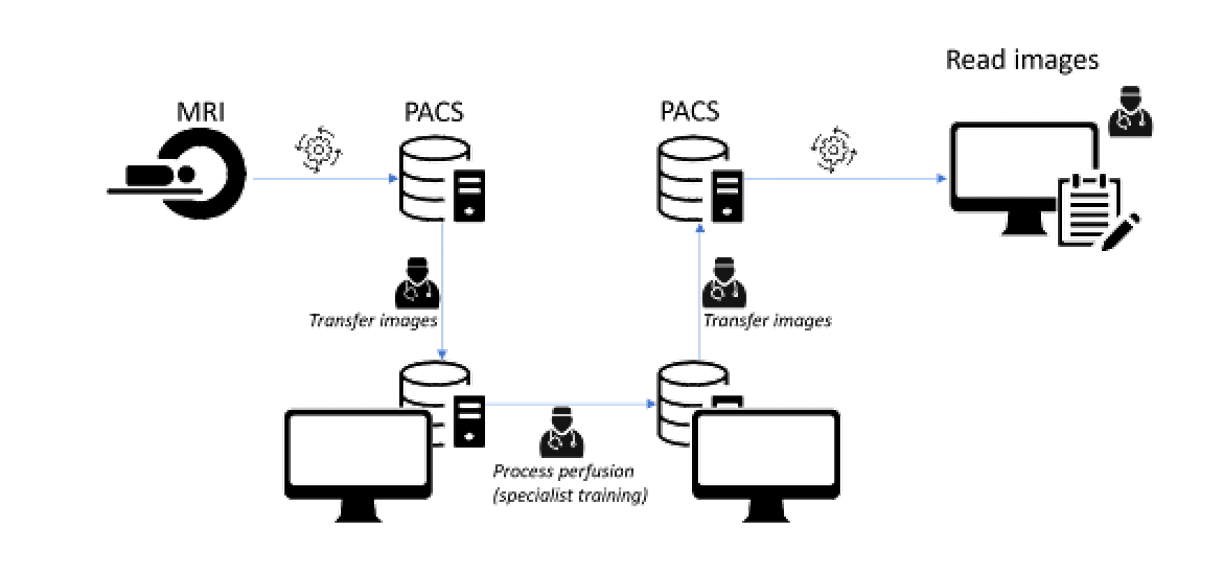
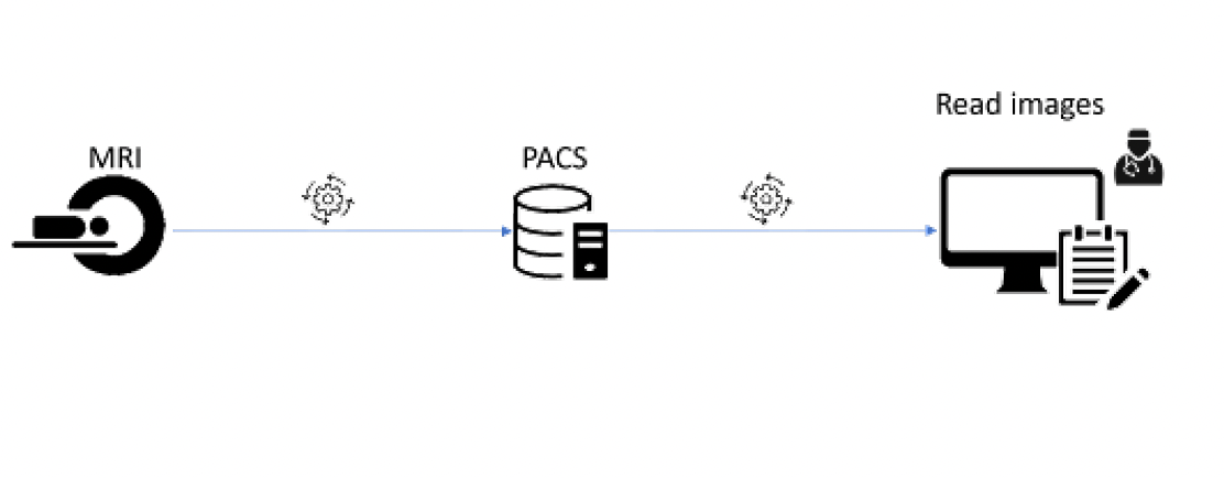
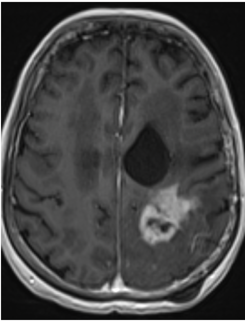
MRI
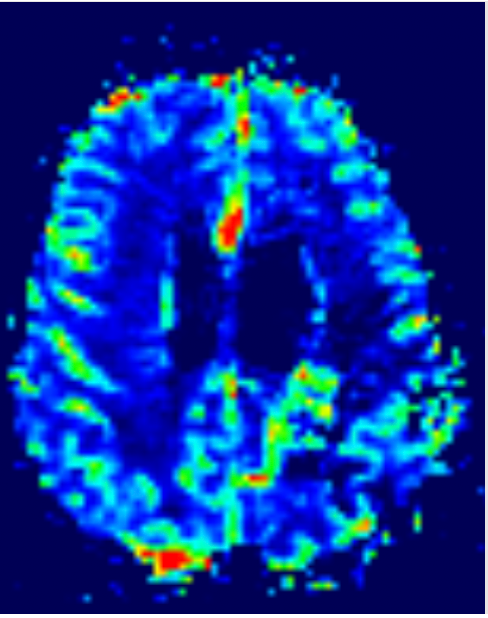
SVD METHOD
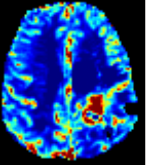
VASCULAR MODEL
Cercare Medical’s Acute Ischemic Stroke modules are designed to enhance the diagnosis and treatment planning for patients with acute ischemic stroke by providing comprehensive and automated analysis of MRI and CT data. Note that some features, such as AI-based detection of lesions, are not available in all jurisdictions.
Fully Automated Processing
Efficiency and Precision: Automatically detects and processes MRI and CT data, ensuring rapid and reliable results. This automation minimizes manual intervention, reducing the risk of human error and streamlining clinical workflows.
Detection of Acute Lesions
Critical Insights: Identifies tissue reflective of ischemic core and hypoperfusion, providing essential information for assessing the extent of brain tissue damage and ischemia. This includes mismatch lesions, volumes, and ratios, which are crucial for treatment planning.
Threshold and AI-Based Methodologies (AI-based detection of lesions, are not available in all jurisdictions.)
Advanced Analysis: Utilizes both threshold-based and AI-based methodologies to enhance the accuracy and reliability of stroke assessment. This dual approach ensures that clinicians have the most accurate information available for decision-making.
Comprehensive Reporting
Streamlined Workflow: Automatically reports results to PACS and/or email notifications to critical personnel, thereby facilitating seamless integration into clinical workflows and ensuring that critical information is readily accessible to the entire care team.
By integrating these advanced features, Cercare Medical’s Acute Ischemic Stroke modules empower clinicians with the tools needed for precise and comprehensive stroke analysis, ultimately aiming at improving patient care and outcomes.
Cercare Medical’s Large Vessel Occlusion (LVO) module is designed to enhance the detection and treatment planning for acute ischemic stroke by providing detailed analysis of CTA data to identify large vessel occlusions.
Fully Automated Processing
Efficiency and Precision: Automatically detects and processes CTA data, ensuring rapid and reliable identification of large vessel occlusions. This automation minimizes the need for manual intervention, reducing the risk of human error and streamlining clinical workflows.
Advanced Detection Algorithms
Critical Insights: Utilizes advanced detection algorithms to accurately identify large vessel occlusions. This is crucial for timely intervention and treatment planning, potentially improving patient outcomes in acute ischemic stroke.
Comprehensive Visualization
Detailed Analysis: Provides comprehensive visualization of vascular structures and occlusions aiming at assisting the clinician in acute image reading to facilitate the assessment and decision-making processes. This includes axial, coronal, and sagittal projections, as well as rotational maximum intensity projections (MIPs) for an in-depth view.
Seamless Reporting
Streamlined Workflow: Automatically reports results to PACS and/or email to critical care personnel, ensuring that critical information is readily accessible to the entire care team and facilitating seamless integration into clinical workflows.
By integrating these advanced features, Cercare Medical’s Large Vessel Occlusion (LVO) module empowers clinicians with the tools needed for precise and comprehensive analysis of large vessel occlusions, with the ultimate goal of improving patient care and outcomes.
Cercare Medical’s Intracerebral Hemorrhage (ICH) module is designed to enhance the detection and assessment of intracerebral hemorrhage using non-contrast CT data, aiding in the diagnosis and treatment planning for patients with hemorrhagic stroke.
Fully Automated Processing
Efficiency and Precision: Automatically detects and processes non-contrast CT data to identify areas of hemorrhage. This automation ensures rapid and reliable results, minimizing manual intervention and reducing the risk of human error.
Advanced Hemorrhage Detection
Critical Insights: Utilizes sophisticated algorithms including AI-based methodology to accurately detect hemorrhagic regions within the brain. This includes detailed mapping of the hemorrhage extent, which is crucial for clinical assessment and treatment planning.
Comprehensive Visualization
Detailed Analysis: Provides detailed visualizations of hemorrhagic regions, including montage images that show every slice of the hemorrhage series in a single 2D image. This comprehensive view, in combination with automated volumetric analysis, aids clinicians in assessing the severity and extent of the hemorrhage.
Automatic Reporting
Streamlined Workflow: Automatically reports results to PACS and/or notifies critical personnel via email, ensuring that critical information is readily accessible to the entire care team. This seamless integration into clinical workflows facilitates efficient and timely decision-making.
By integrating these advanced features, Cercare Medical’s Intracerebral Hemorrhage (ICH) module empowers clinicians with the tools needed for precise and comprehensive hemorrhage analysis, ultimately aimed at improving patient care and outcomes.
Cercare Medical’s ASPECTS module is designed to enhance the detection and assessment of early signs of ischemia using non-contrast CT data, aiding in the diagnosis and treatment planning for patients with hemorrhagic stroke.
Fully Automated Processing
Efficiency and Precision: Automatically detects and processes non-contrast CT data to identify brain areas affected by ischemia. This automation ensures rapid and reliable results, minimizing manual intervention and reducing the risk of human error.
Advanced ASPECT Scoring
Critical Insights: Utilizes sophisticated algorithms to accurately detect early signs of ischemia in the classical ASPECT regions. This includes detailed mapping of the ASPECT regions in addition to the standard single-image overview, which provides additional options for clinical inspection and treatment planning.
Comprehensive Visualization
Detailed Analysis: Provides detailed visualizations of ASPECTS-based early signs of ischemia detection, including summary images that show the standard ASPECT regions alongside the ASPECT score in a single 2D image. This summary view in essence reduces an extensive non-contrast CT series to a single image, thus aiding clinicians in assessing the extent of severe ischemia.
Automatic Reporting
Streamlined Workflow: Automatically reports results to PACS and/or notifies critical personnel via email, ensuring that critical information is readily accessible to the entire care team. This seamless integration into clinical workflows facilitates efficient and timely decision-making.
By integrating these advanced features, Cercare Medical’s ASPECT capabilities empowers clinicians with the tools needed for precise and comprehensive analysis of early signs of ischemia, ultimately aimed at improving patient care and outcomes.
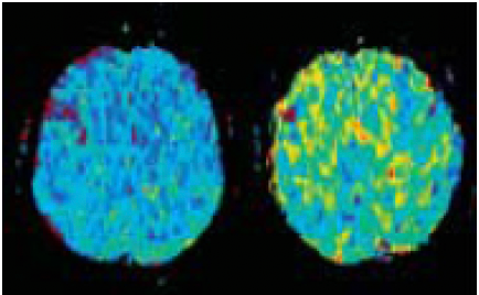
MTT

MTT

T2 FLAIR
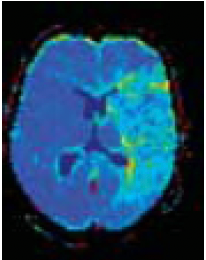
MTT

MTT
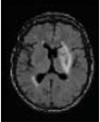
T2 FLAIR

