Biased visualization of hypoperfused tissue by computed tomography due to short imaging duration: improved classification by image down-sampling and vascular models.

Mikkelsen IK, Jones PS, Ribe LR, et al. Eur Radiol. 2015;25(7):2080-2088

Abstract

Objectives

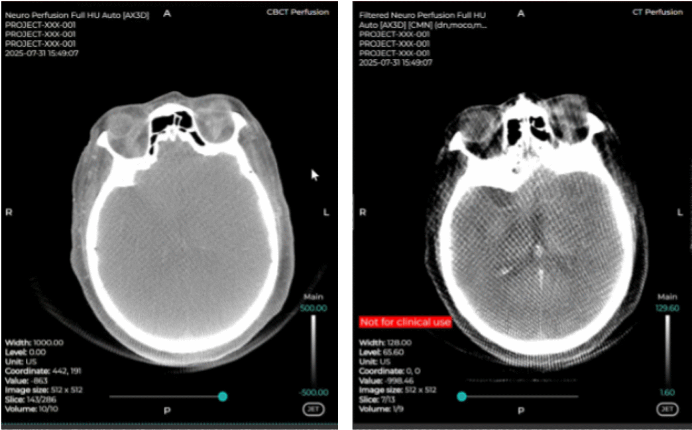
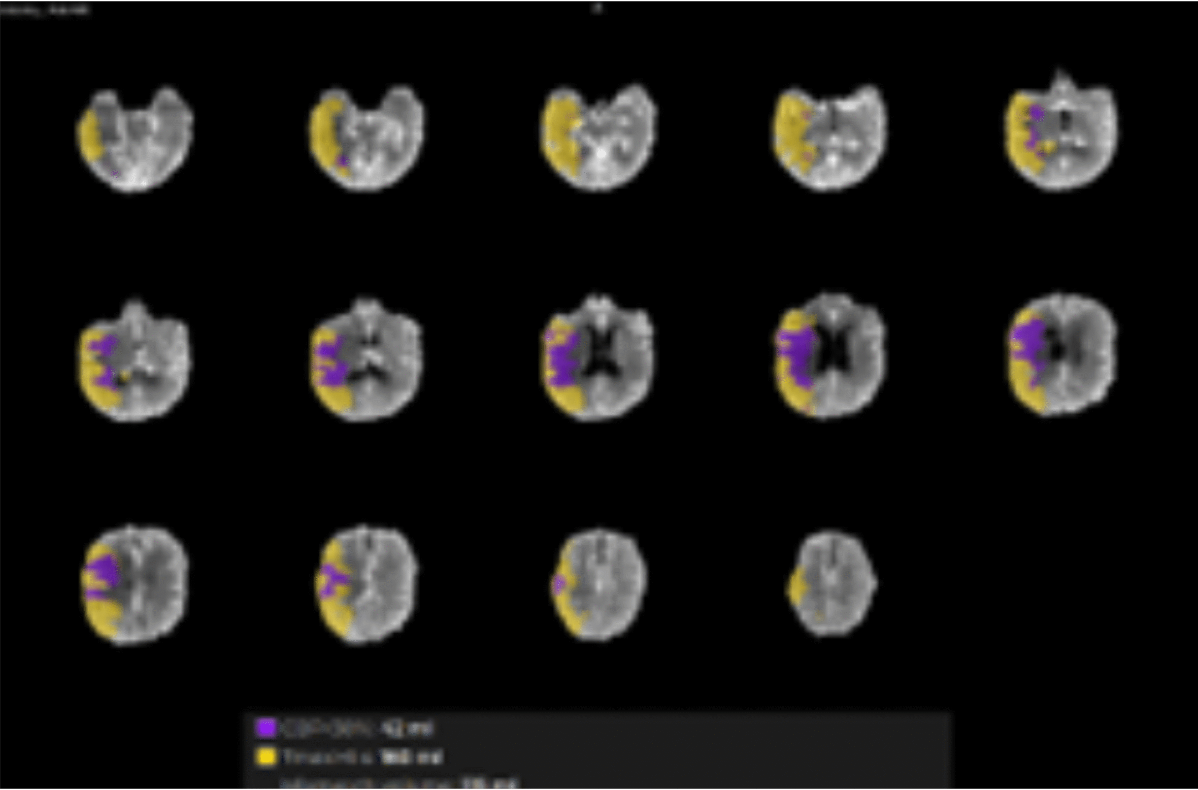
Lesion detection in acute stroke by computed-tomography perfusion (CTP) can be affected by incomplete bolus coverage in veins and hypoperfused tissue, so-called bolus truncation (BT), and low contrast-to-noise ratio (CNR). We examined the BT-frequency and hypothesized that image down-sampling and a vascular model (VM) for perfusion calculation would improve normo- and hypoperfused tissue classification.

Conclusion

BT is not uncommon in stroke CTP with 40-s scan duration. Applying image down-sampling and VM improve tissue classification.

Get access to the full article: doi:10.1007/s00330-015-3602-x

Ready to Transform Your Diagnostic Capabilities?

Join Us in Advancing Brain Health with Cutting-Edge Microvascular Modeling.
Contact Us Today to Learn More!
© 2026 Cercare Medical. All rights reserved.

CLINICAL USE CASES AND TECHNICAL HIGHLIGHTS


CLINICAL USE CASES
  • Directly impacts the patient treatment pathway, both intra- and post-intervention.
  • Assess outcomes post-intervention: detect missed stenoses, distal occlusions, or microvascular disturbances.
  • Guide complementary therapies on the spot based on perfusion status.
  • May detect the no-reflow phenomenon — providing insight into cases where recovery may fail despite successful thrombectomy.
  • Potentially enables direct-to-angio workflows.

TECHNICAL HIGHLIGHTS
  • Full set of perfusion markers: CBF, CBV, MTT.
  • Advanced oxygen metabolism imaging: OEF, CMRO₂.
  • Most reliable results with 10 CBCT acquisitions.
  • Vendor-agnostic platform.

CBCT Imaging

Side-by-side comparison: The left-hand side is the raw CBCT data, while the right-hand side panel is the pre-processed data.
Using CMN software in the angio suite delivers results comparable to conventional CT/MR perfusion.

Explore CMN!

Fast. Reliable. Personalized.

Discover how Cercare Medical Neurosuite (CMN) enhances post-processing images and provides actionable insights from advanced biomarkers - tailored to your workflow.

Fill-out the form below, and a representative will contact you.

Thank you for uploading your DICOM files.

Your submission has been received successfully.

Next steps:

  • Our team will analyze the uploaded data.
  • A Cercare Medical representative will reach out to you once the analysis is complete.
  • Rest assured that all files will be deleted immediately after use, in line with our data protection policies.

Sign up for the upcoming Perfusion & Metabolic Imaging Community (PMIC)

Request a demo

Let us give you a tour around our solution.

Fill in your credentials, and a member of the Cercare Medical team will contact you with a date and time proposal that fits your schedule.
})