If you are a driven professional with a passion for innovation and excellence, we want to hear from you. Explore exciting career opportunities and become a part of our mission to revolutionize medical imaging. Join Cercare Medical and help us make a real impact on the healthcare industry.
Ready to Transform Your Diagnostic Capabilities?
Join Us in Advancing Brain Health with Cutting-Edge Microvascular Modeling. Contact Us Today to Learn More!
Guide complementary therapies on the spot based on perfusion status.
May detect the no-reflow phenomenon — providing insight into cases where recovery may fail despite successful thrombectomy.
Potentially enables direct-to-angio workflows.
TECHNICAL HIGHLIGHTS
Full set of perfusion markers: CBF, CBV, MTT.
Advanced oxygen metabolism imaging: OEF, CMRO₂.
Most reliable results with 10 CBCT acquisitions.
Vendor-agnostic platform.
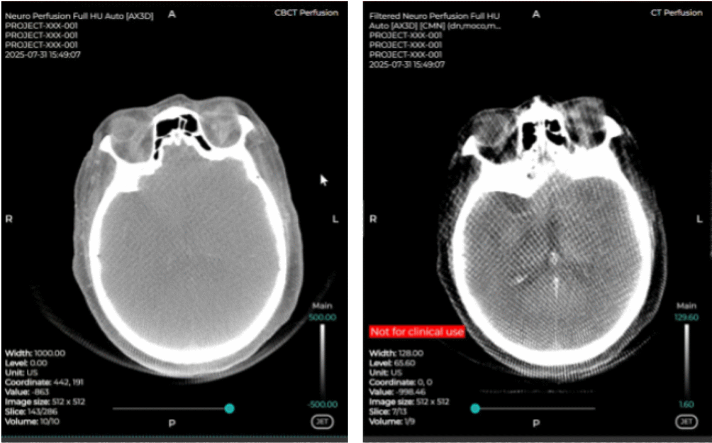
CBCT Imaging
Side-by-side comparison: The left-hand side is the raw CBCT data, while the right-hand side panel is the pre-processed data.
Using CMN software in the angio suite delivers results comparable to conventional CT/MR perfusion.
Explore CMN!
Fast. Reliable. Personalized.
Discover how Cercare Medical Neurosuite (CMN) enhances post-processing images and provides actionable insights from advanced biomarkers - tailored to your workflow.
Fill-out the form below, and a representative will contact you.
Thank you for uploading your DICOM files.
Your submission has been received successfully.
Next steps:
Our team will analyze the uploaded data.
A Cercare Medical representative will reach out to you once the analysis is complete.
Rest assured that all files will be deleted immediately after use, in line with our data protection policies.
Sign up for the upcoming Perfusion & Metabolic Imaging Community (PMIC)
Imaging
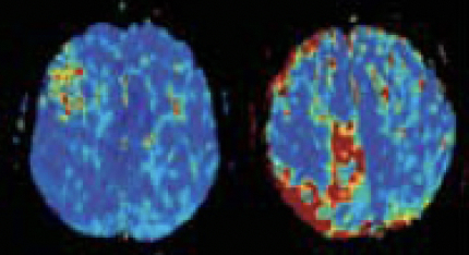
MTT
SVD model
MTT
Vascular Model
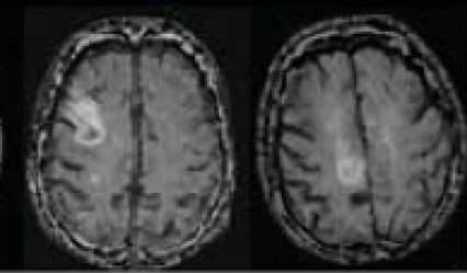
T2 FLAIR
follow-up image
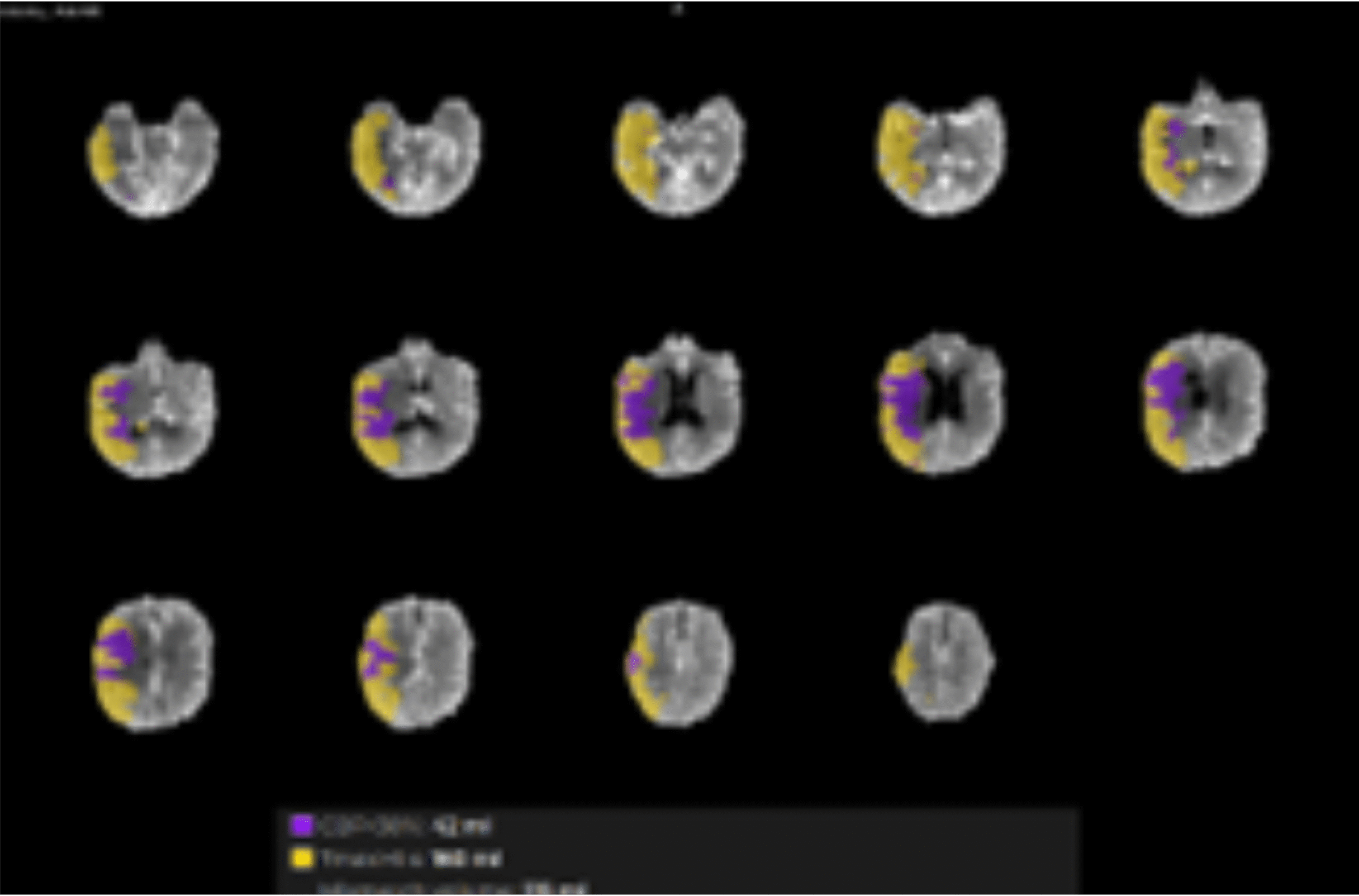
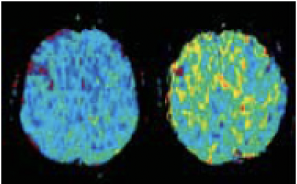
Perfusion maps of a stroke patient show a large difference in lesion appearance compared to the SVD technique, where the VM appears in better correspondence with the T2 FLAIR follow-up.
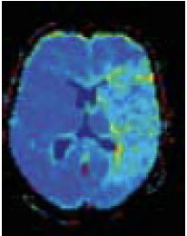
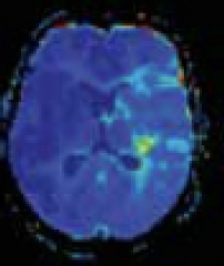
MTT
SVD model
MTT
Vascular Model
T2 FLAIR
follow-up image
The modest degree of tissue involvement is consistent with the moderate neurologic deficits, National Institutes of Health Stroke Scale (NIHSS) = 4.
Request a demo
Let us give you a tour around our solution.
Fill in your credentials, and a member of the Cercare Medical team will contact you with a date and time proposal that fits your schedule.
Your application has been successfully submitted.
Our recruitment team will review it and contact you if there is a next step.