Improved Co-Registration: Enhancements ensure better co-registration for cases with significant rotation and thin-slice data, improving overall accuracy.
Enhanced M1-M6 Region Appearance: The M1-M6 regions now display without non-linear warping artifacts, ensuring consistent and accurate representation across subjects.
New ASPECTS Overview Montage: Introduces a montage featuring only two slices of the brain, available with and without the ASPECTS overlay. The montage also includes a table with intensity information, highlighting affected regions and providing the ASPECT scores for both hemispheres.
ICH Dependency Warning: A warning is now included in ASPECTS outputs if the ICH module detects bleeding. This guides the user to verify suspected bleeding and ensures ASPECTS is not used if bleeding is confirmed.
ASPECT Score in CMN DICOM Header: The ASPECT score is now added to the CMN DICOM header for easier reference and integration.
Fused Series: Combines any two series with adjustable overlay opacity, allowing for seamless export to PACS.
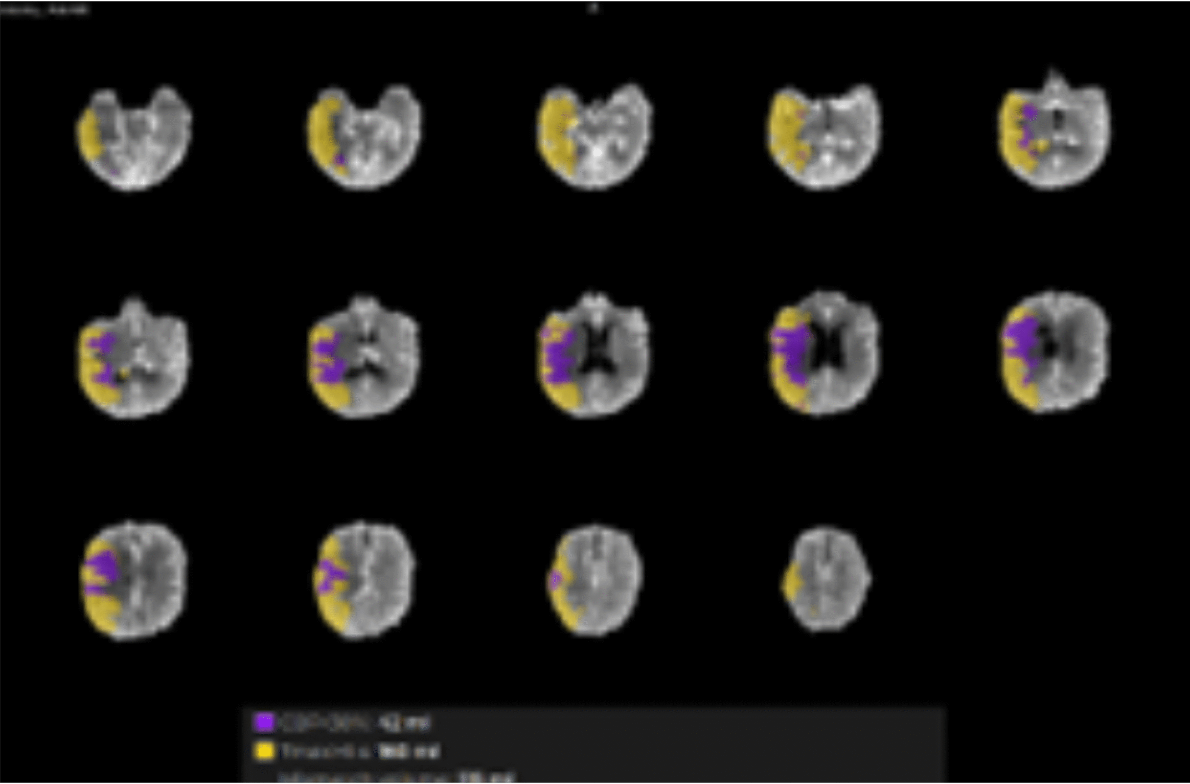
Montages: Displays the lesion overlaid on a series of anatomical images in a single image, incorporating all relevant quantitative lesion data.
Overlay Series: Overlays the lesion on a series of anatomical images in a scrollable format, enabling detailed analysis with all relevant quantitative lesion data.
Supports analysis of diseases like Moyamoya by processing multiple series of the same sequence time (e.g., pre- and post-treatment DSC series).
Ensures distinction between derived series using Acquisition Date Time.
Optimization across workflows for faster processing.
Enables advanced evaluation of wake-up strokes by identifying DWI/FLAIR mismatch, helping assess if the tissue is ischemic but salvageable.
Comprehensive Longitudinal Analysis: CMN enables longitudinal analysis by comparing data from multiple studies of a patient. This includes insights into both lesion growth and perfusion changes over time.
Automatic VOI Analysis: Facilitates automatic Volume of Interest (VOI) analysis for segmented brain tumor lesions, streamlining research workflows.
Quantitative and Visual Insights: Generates detailed quantitative data and scrollable montages, offering a clear visualization of perfusion changes and lesion growth over time.
Customizable Map Inclusion: Allows the user to include any perfusion or diffusion map of interest in the VOI analysis, ensuring flexibility and relevance to specific research needs.
Introducing a parameter-driven license system that allows mixing clinical and research features in the same license.
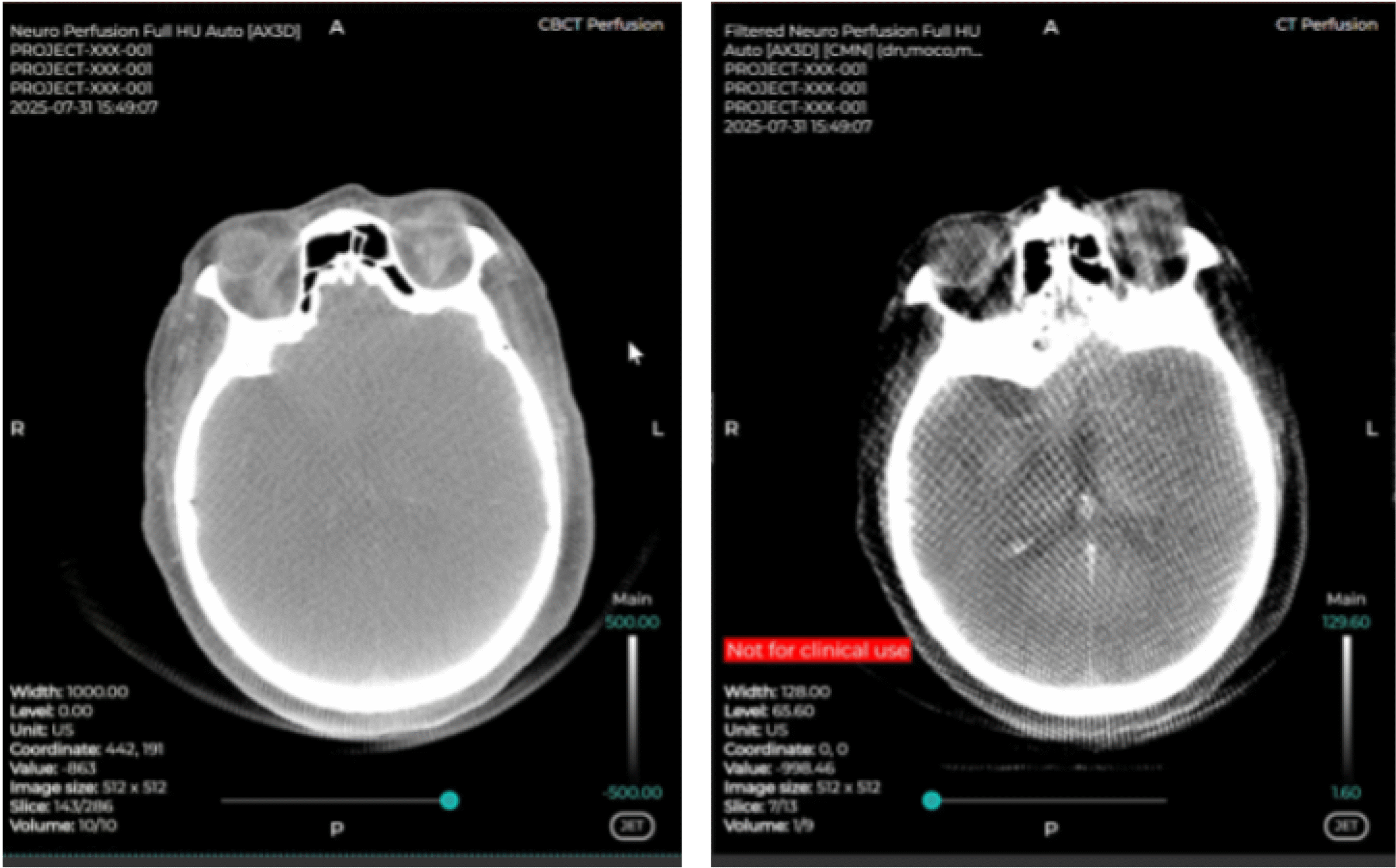
Research outputs are clearly marked “not for clinical use,” enabling seamless use of clinically approved outputs alongside features and outputs intended for research only.