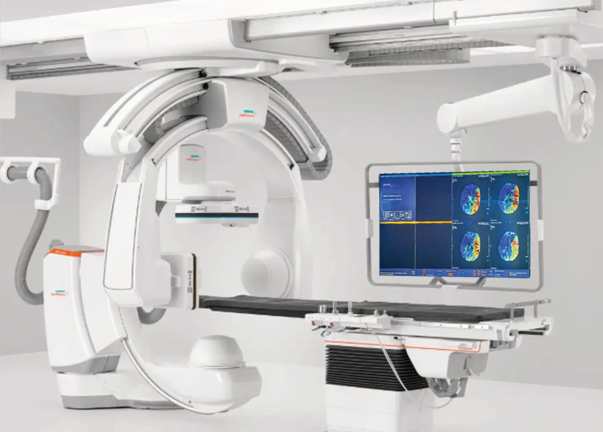
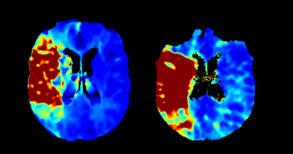
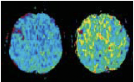
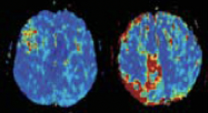
1 in 3 patients show no-reflow despite high mTICI scores after thrombectomy. DSA confirms the vessel is open; it cannot confirm capillary-level reperfusion. Cercare CBCT Perfusion generates CT-quality perfusion maps — rCBF, rCBV, MTT, Tmax, plus CTH, OEF and CMRO₂ — from your existing C-arm. No patient transfer. Real-time tissue assessment while the team is still in the room.
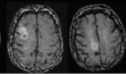
Thrombectomy has transformed acute stroke care. Yet, in some patients, microvascular obstruction - known as the no-reflow phenomenon - prevents full reperfusion, limiting recovery despite technically successful procedures. Detecting this phenomenon can provide crucial insights into treatment outcomes. The missing link is the ability to see beyond the vessel - to assess tissue health and viability in real time.
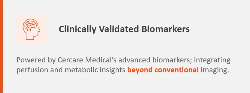
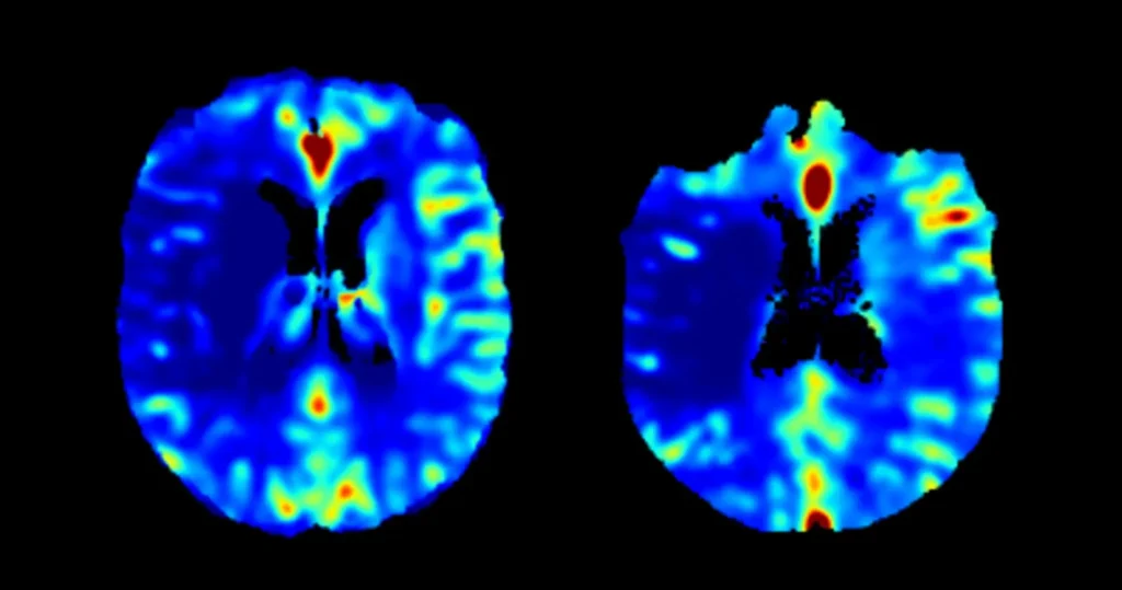
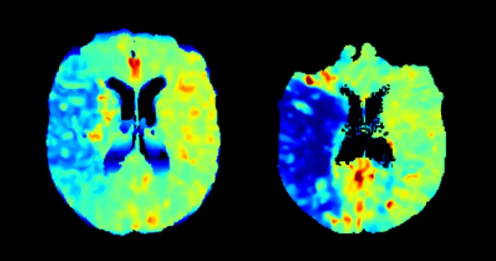
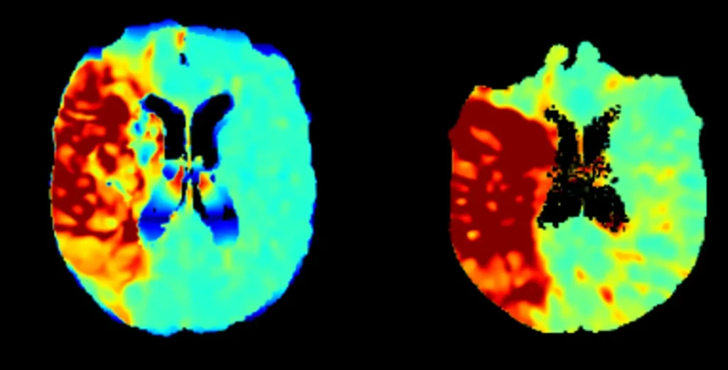
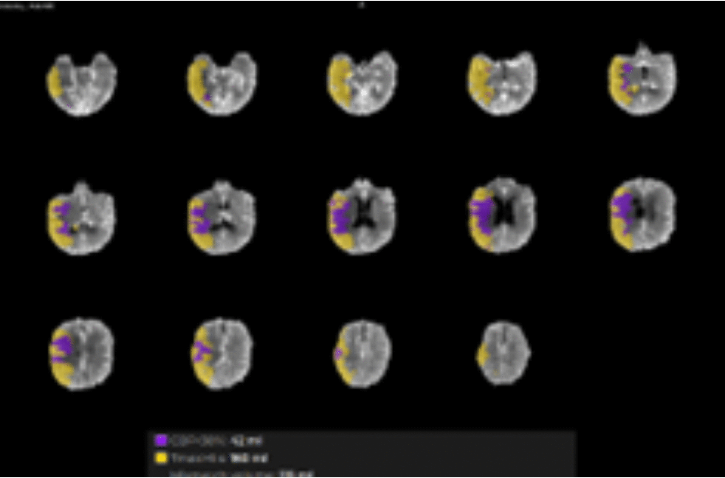
In addition to conventional biomarkers of blood flow and volume, Cercare Medical provides unique insights into tissue metabolism through oxygen-based biomarkers. By combining CTH, OEF, and CMRO₂, we map oxygen availability alongside perfusion, enabling deep understanding of microvascular conditions and supporting more precise clinical interpretation.
This foundation now powers a new breakthrough:
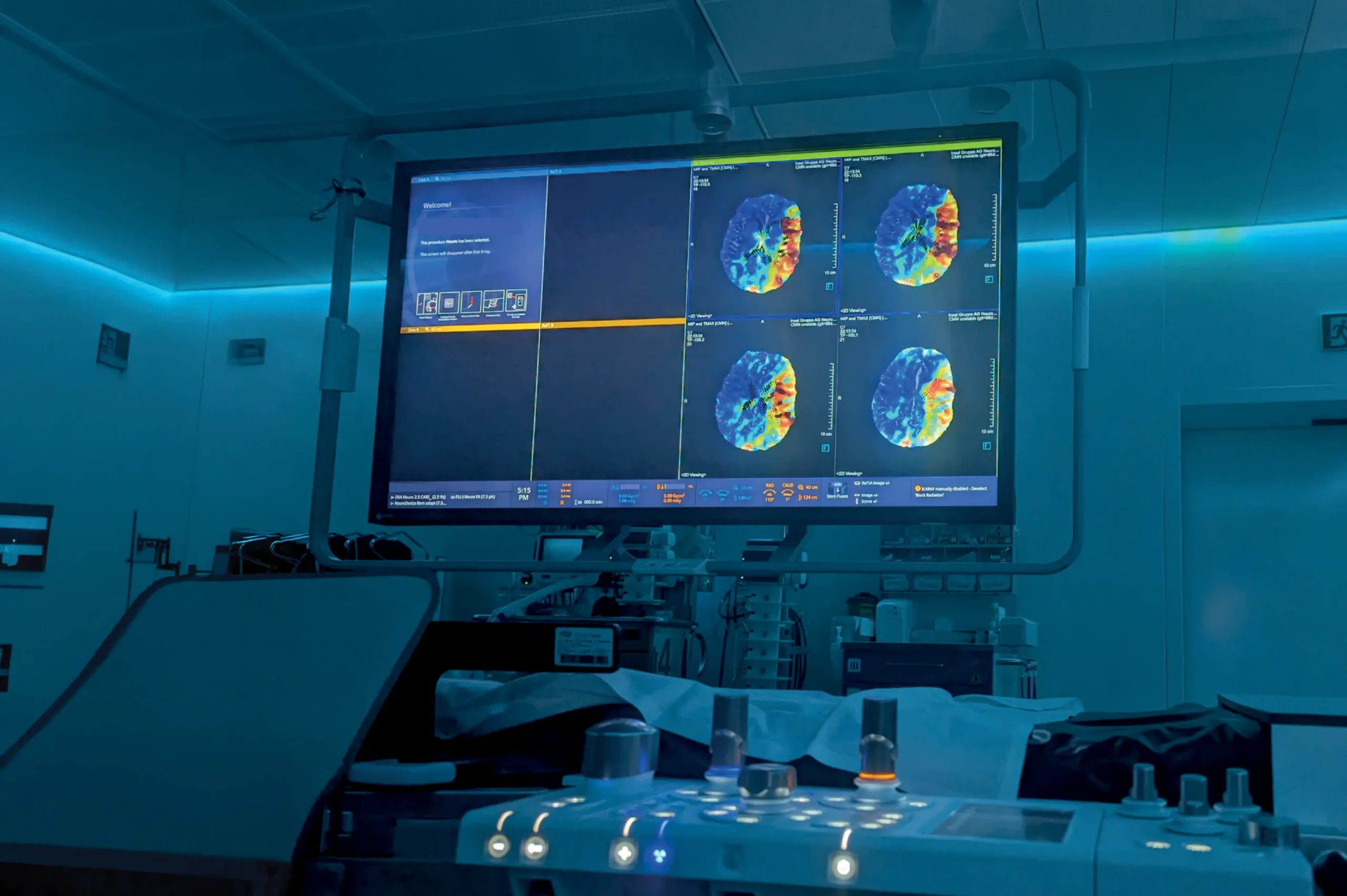
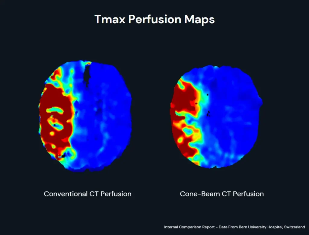
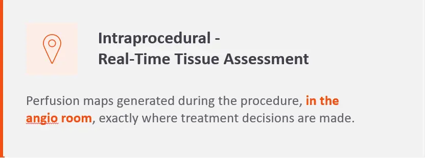
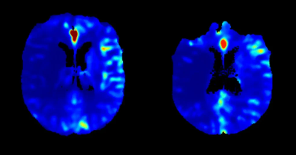
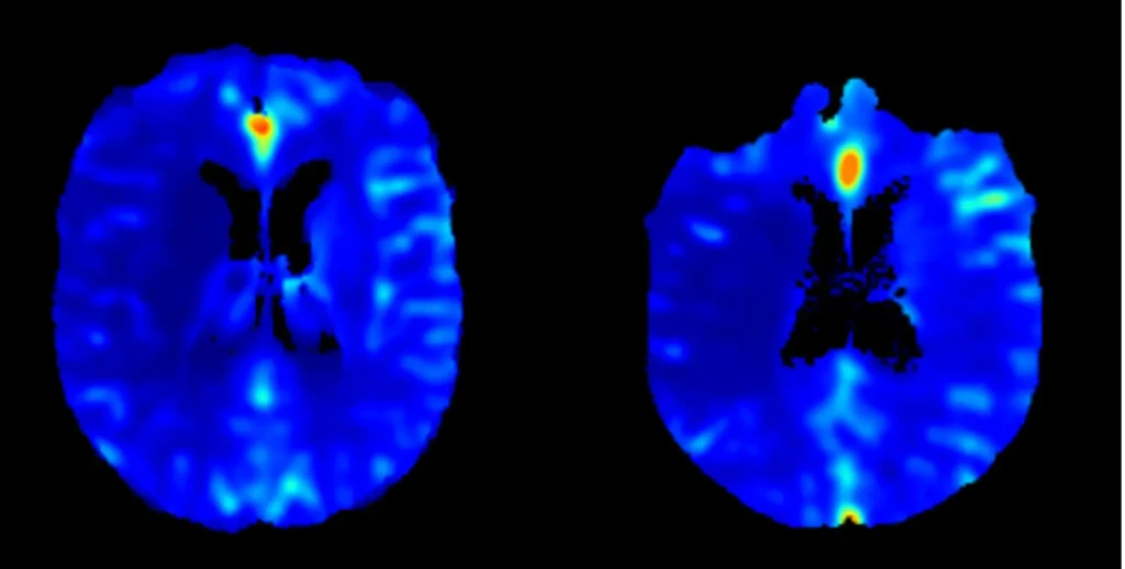
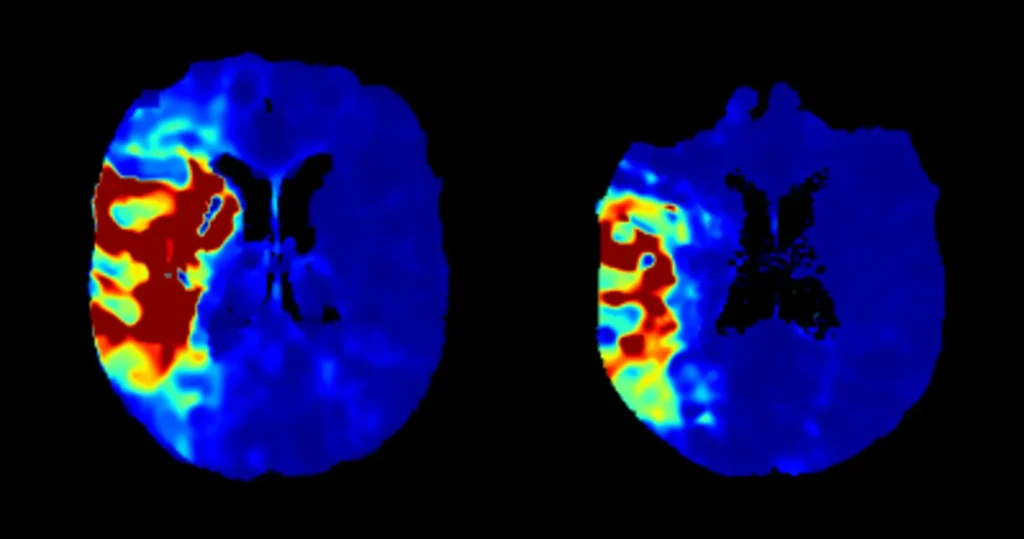
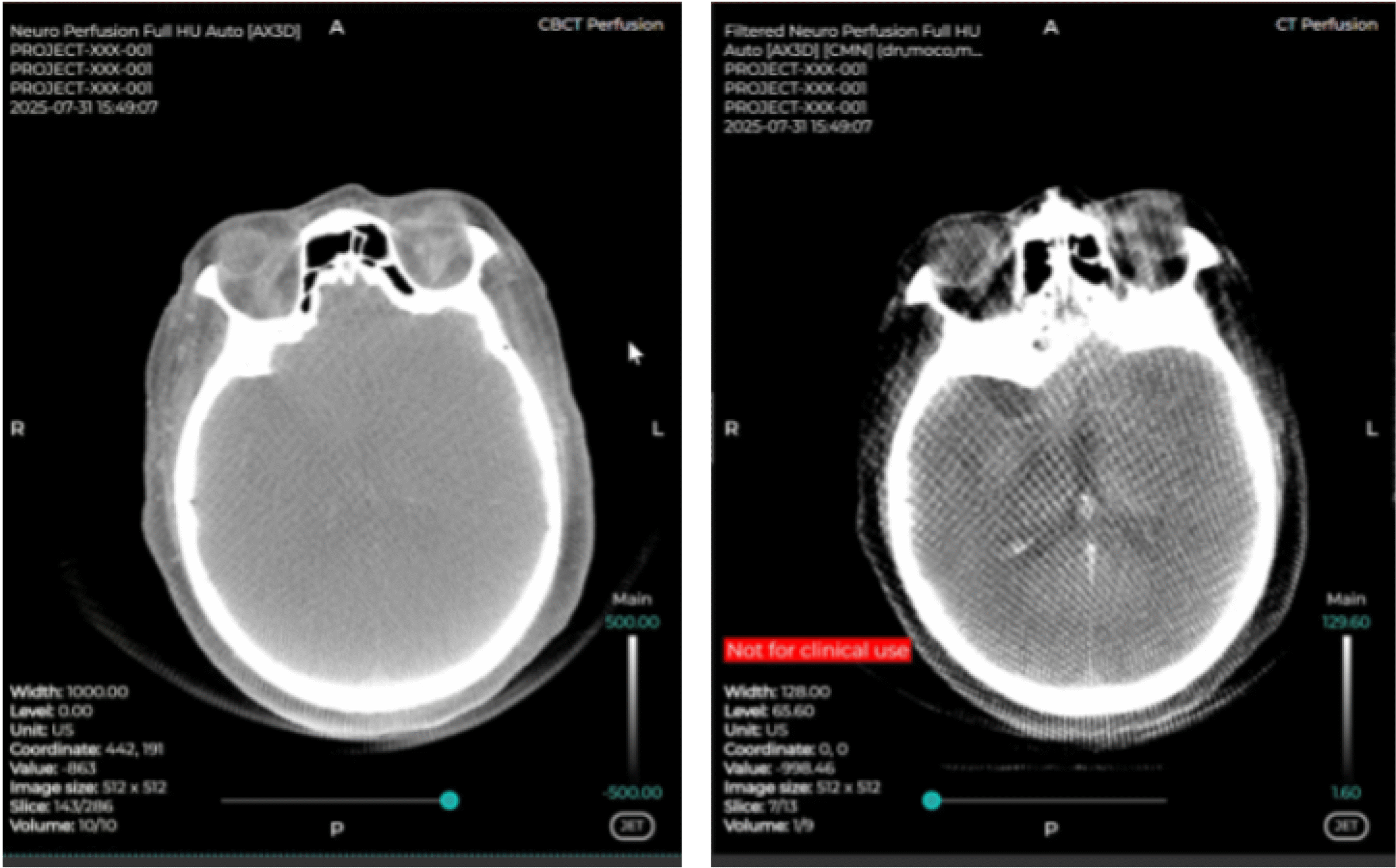
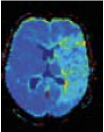
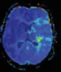
Cone-Beam CT Perfusion which allows INTRAPROCEDURAL perfusion imaging and provides real-time assessment of tissue perfusion before and after thrombectomy, enabling clinicians to evaluate the physiological impact of treatment beyond angiographic vessel reopening.
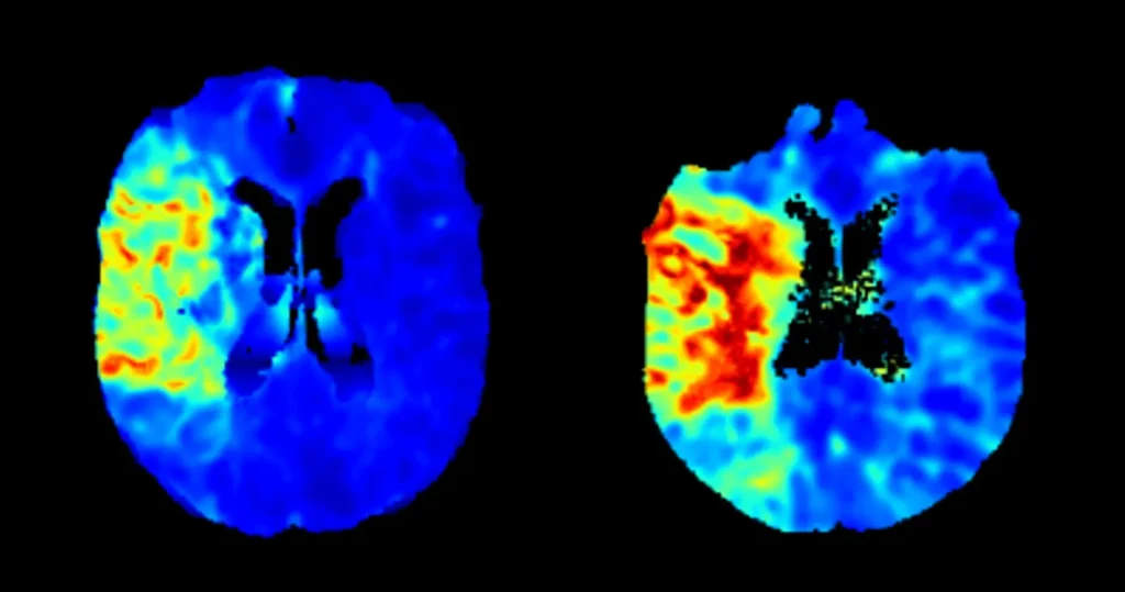
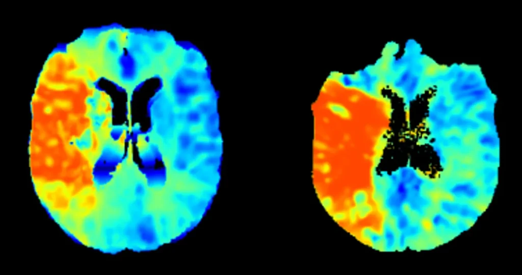
While angiography can confirm successful recanalization of the occluded vessel, it cannot determine whether blood flow has been effectively restored at the microvascular and tissue level. Yet it is microvascular reperfusion that ultimately drives tissue salvage and patient outcome.
For clinicians, Cone-Beam CT Perfusion means objective physiological feedback during the intervention—helping confirm procedural success, reduce uncertainty, and support evidence-based decision-making when every minute matters.