STUDY HIGHLIGHT

Cortical Thickness Changes Precede High Levels of Amyloid by at Least 7 Years
Can Detect Signs of Alzheimer’s Disease Much Earlier Than Thought
How early do brain changes associated with Alzheimer’s disease become detectable with imaging?

Because the biology underlying Alzheimer's disease is thought to unfold over decades - beginning long before cognitive symptoms appear - a major research focus is to identify brain biomarkers of impending disease.
 
It has long been thought that the earliest detectable signal on brain scans in Alzheimer's is amyloid pathology as seen on PET scans.
 
But new research published in Nature Neuroscience suggests that measurable changes in brain structure may occur at least seven years before amyloid pathology becomes detectable with PET. This suggests that harmful processes may already be underway in the brain long before amyloid pathology becomes detectable on imaging.
 
The study followed healthy individuals with regular brain scans (MRI) over nearly two decades, allowing the researchers to look back at brain MRI scans in those who later developed amyloid pathology - acquired years before amyloid became detectable.
 
We are especially proud to highlight that James Michael Roe, Ph.D., International Scientific Lead at Cercare Medical, is the first author of the study, which he led during his time at the University of Oslo and The University of California, Berkeley.
 
The findings provide new insights into how early brain changes associated with Alzheimer’s disease may begin and could contribute to a better understanding of the brain processes taking place long before cognitive symptoms appear. 

At Cercare Medical, we take pride in our colleagues' contributions to science.

This study has already had a major impact, having been read more than 18,000 times in its first month.
>>> NeuroScience: Cortical Thickness Changes Precede High Levels of Amyloid by at Least 7 Years >>> University of Oslo: Can detect signs of Alzheimer's disease much earlier than thought

About Cercare Medical
Cercare Medical is a provider of advanced perfusion imaging solutions. With fully automated, vendor-neutral software for CT, CBCT, and MRI perfusion post-processing, the company supports clinicians across the world with tools that enhance diagnostics for stroke, oncology, Alzheimer’s disease, and neurological conditions. Cercare’s proprietary biomarkers and automated pipelines enable better imaging maps, improving speed and confidence in clinical decision-making.

Ready to Transform Your Diagnostic Capabilities?

Join Us in Advancing Brain Health with Cutting-Edge Microvascular Modeling.
Contact Us Today to Learn More!
© 2026 Cercare Medical. All rights reserved.

CLINICAL USE CASES AND TECHNICAL HIGHLIGHTS


CLINICAL USE CASES
  • Directly impacts the patient treatment pathway, both intra- and post-intervention.
  • Assess outcomes post-intervention: detect missed stenoses, distal occlusions, or microvascular disturbances.
  • Guide complementary therapies on the spot based on perfusion status.
  • May detect the no-reflow phenomenon — providing insight into cases where recovery may fail despite successful thrombectomy.
  • Potentially enables direct-to-angio workflows.

TECHNICAL HIGHLIGHTS
  • Full set of perfusion markers: CBF, CBV, MTT.
  • Advanced oxygen metabolism imaging: OEF, CMROâ‚‚.
  • Most reliable results with 10 CBCT acquisitions.
  • Vendor-agnostic platform.

CBCT Imaging

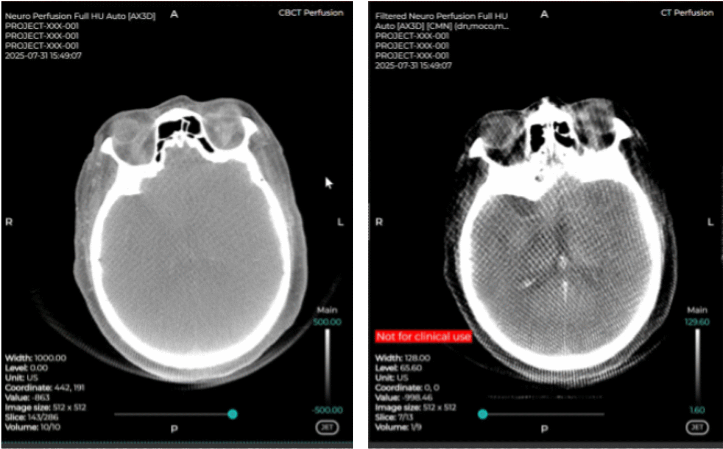
Side-by-side comparison: The left-hand side is the raw CBCT data, while the right-hand side panel is the pre-processed data.
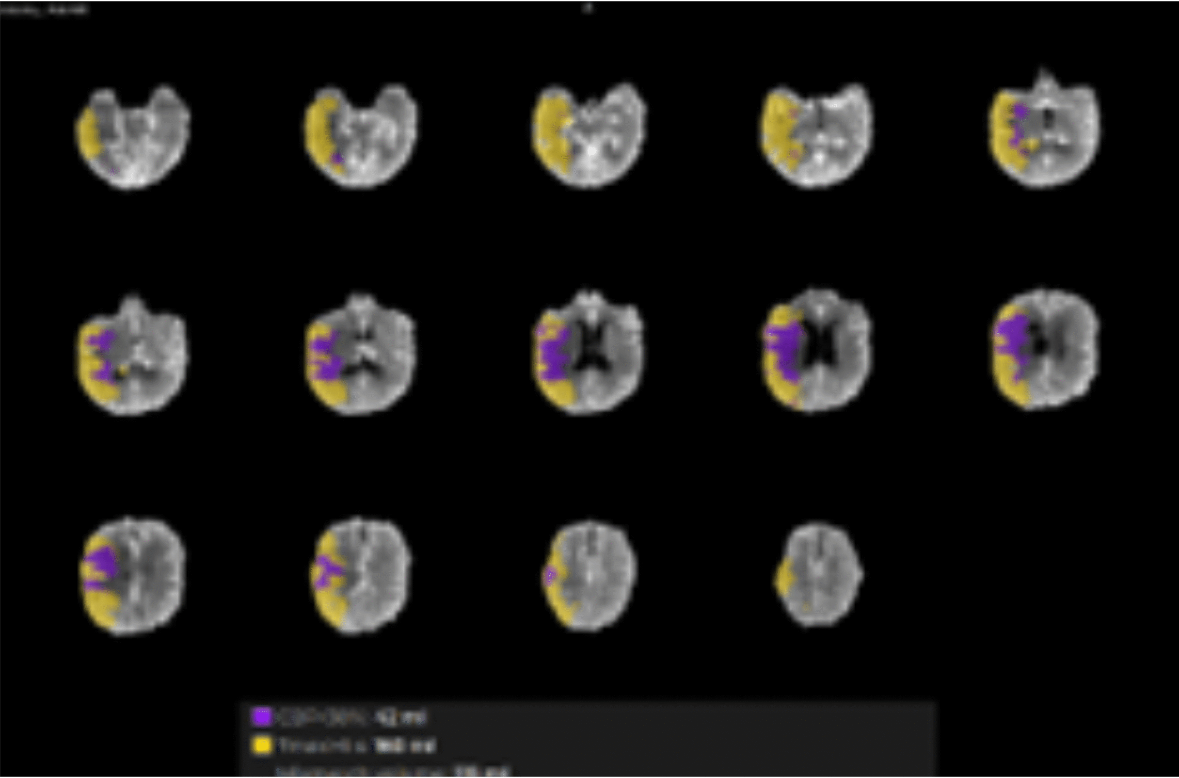
Using CMN software in the angio suite delivers results comparable to conventional CT/MR perfusion.

Contact us:

A Cercare Medical Representative will reach out to you directly to assist with your inquiry.

Sign up for the upcoming Perfusion & Metabolic Imaging Community (PMIC)

Request a Demo

Let us give you a tour around our solution.

Fill in your credentials, and a member of the Cercare Medical team will contact you with a date and time proposal that fits your schedule.

Explore CMN!

Fast. Reliable. Personalized.

Discover how Cercare Medical Neurosuite (CMN) enhances post-processing images and provides actionable insights from advanced biomarkers - tailored to your workflow.

Fill-out the form below, and a representative will contact you.

Thank you for uploading your DICOM files.

Your submission has been received successfully.

Next steps:

  • Our team will analyze the uploaded data.
  • A Cercare Medical representative will reach out to you once the analysis is complete.
  • Rest assured that all files will be deleted immediately after use, in line with our data protection policies.
})